Galerie
Slide shows
| Differenziertes Krafttraining | |
| Blum: Kinesiologische Analyse | |
| Unterkiefer | |
| Ren mobilis (Die Wanderniere) |
Schriftliches

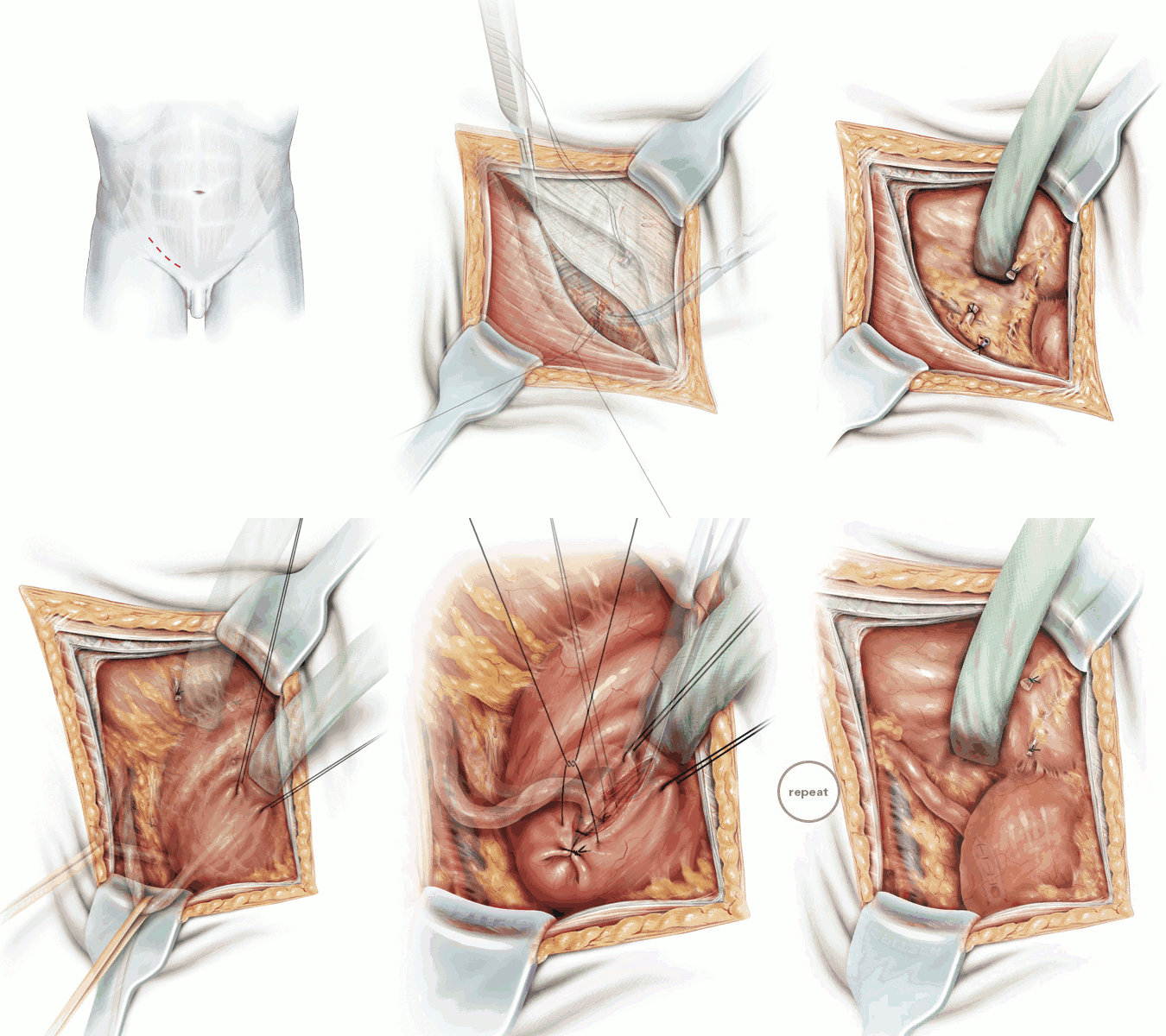
| Title | Antireflux surgery: Lich-Gregoir extravesical ureteric tunneling |
| Technique | Pencil / Watercolour / Computer |
| Author | Riedmiller/Gerharz, Univerity medical School Wuerzburg |
| Publication | British Journal of Urology International (BJUI) |
| Publisher | Blackwell-Wiley Publishing 2008 |
| close window | |
| Figure 1 | Oblique incision in the right lower quadrant (Gibson) |
| Figure 2 | Division of the external oblique aponeurosis in the direction of the fibres |
| Figure 3 | Separation of the internal oblique muscle and the transversalis abdominis muscle |
| Figure 4 | Ligation and division of the epigastric vessels |
| Figure 5 | Division of the thin transversalis fascia |
| Figure 6 | Dissection of the spermatic chord, craniomedial mobilization of the peritoneum, opening of the subperitoneal space (iliac vessels exposed) |
| Figure 7 | Dissection and division of the hypogastric vessels which are crossing the ureter |
| Figure 8 | Mobilization of the ureter, encircling with a surgical loop |
| Figure 9 | Stay sutures help to rotate the bladder medially, incision follows the natural course of the ureter |
| Figure 10 | Vertical division of the detrusor muscle. Y-shaped incision proximally to release the flaps |
| Figure 11 | Placing the ureter in the groove in contact with the bladder epithelium, loose closure of the muscle over the ureter with interrupted sutures |
| Figure 12 | An Overhold clamp is used to ensure a sufficiently wide entry of the tunnel |
| Figure 13 | Allowing the bladder to fall back and resume its physiological position |
| close window | |