Galerie
Slide shows
| Differenziertes Krafttraining | |
| Blum: Kinesiologische Analyse | |
| Unterkiefer | |
| Ren mobilis (Die Wanderniere) |
Schriftliches

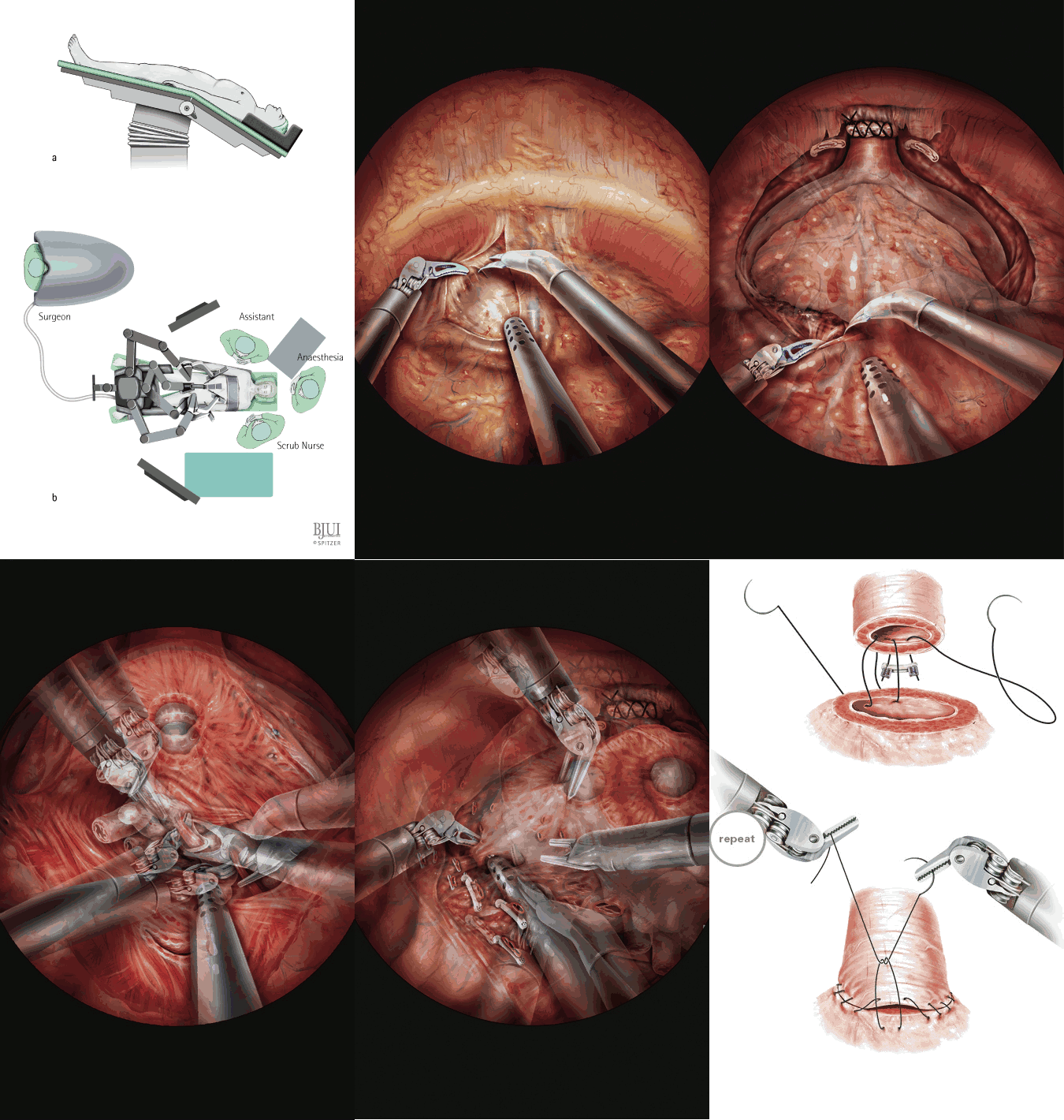
| Title | Robot-assisted laparoscopic nerve-sparing prostatectomy |
| Technique | Pencil / Watercolour / Computer |
| Author | Gillitzer/Thueroff et al., University Medical Center Mainz |
| Publication | British Journal of Urology International (BJUI) |
| Publisher | Blackwell-Wiley Publishing 2009 |
| close window | |
| Figure 1 | a) Specific Patient positioning b) Surgical team |
| Figure 2 | Port placement |
| Figure 3 | The bladder is mobilized from the anterior abdominal wall |
| Figure 4 | The visceral layer of the endopelvic fascia is superficially incised |
| Figure 5 | The levator muscle and the puboperinealis muscle are dissected and swept laterally |
| Figure 6 | The tissue overlying the urethra is transected while the assistant pushes the prostate down |
| Figure 7 | The dorsal vein complex is oversewn |
| Figure 8 | The periprostatic fascia is bluntly and sharply dissected from the prostatic capsule |
| Figure 9 | Dissection of the bladder neck can be started |
| Figure 10 | The ventral circumference of the bladder neck has been devided, The Foley catheter is deflated |
| Figure 11 | The tip of the catheter is pulled ventrally, opening of the retrotrigonal space |
| Figure 12 | The fourth roboter arm elevates the vas deferens |
| Figure 13 | The vasa have been clipped and transected. Dissection of the seminal vessels |
| Figure 14 | Clipping and transecting of the vessels between the lobes of the seminal vesicles |
| Figure 15 | Denonvilliers' fascia is cut transversally |
| Figure 16 | Separation of periprostatic fascia with the NVBs |
| Figure 17 | Management of the prostatic pedicles |
| Figure 18 | Dissection towards the apex of the prostata |
| Figure 19 | Ventral circumference of the urethra is incised |
| Figure 20 | The rectourethralis muscle is readapted to the transected retrotrigonal muscle layer |
| Figure 21 | The vesicourethral anastomosis is completed |
| close window | |